Cerebellar Imaging and Ataxias
Our research focuses on cerebellar imaging and digital biomarkers in neurodegeneration – with particular emphasis on ataxias. We aim to improve diagnosis and monitor disease progression in ataxia and related movement disorders.
|

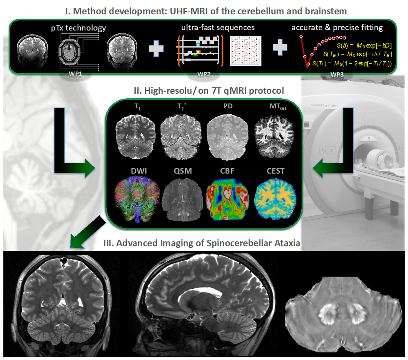
Ultra Highfield MRI
Ultra-high magnetic fields (UHF) offer significantly higher resolutions and advanced MR applications, such as iron-sensitive sequences and metabolic imaging. Thus, UHF-MR has an enormous potential to detect and monitor structural and chemical alterations at a. level of detail that cannot be achieved with a conventional fieldstrength of 3T. However, these clear advantages are opposed by technical shortcommings of field inhomogeneities, which fundamentally degrade image quality, particularly at the excentric infratentorial regions of the field of view. In collaboration with the MR Physics group at the DZNE, lead by Tony Stöcker, we have successfully implemented a comprehensive protocol including structural and metabolic sequences (T1w, T2w, FLAIR, MPM, QSM, CEST) using parallel transmission technologies (pTx) that allows imaging of the entire brain including brainstem and cerebellum in consistently high resolution without artifacts at a fieldstrength of 7T. The acquisition in patients suffering from movement disorders is currently ongoing. We hypothesize, that the increased sensitivity of UHF-MRI has the potential to substantially improve the understanding of structural and metabolic brain alterations in neurodegenerative diseases.
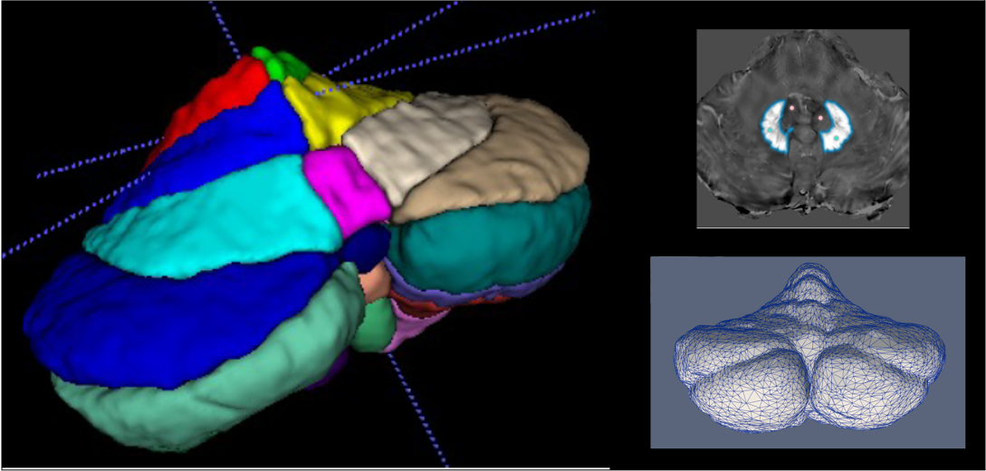
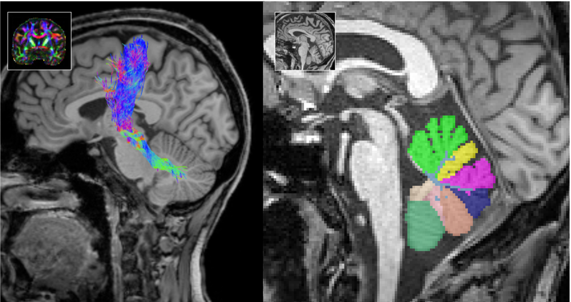
Automated Segmentations & AI-driven MRI Analysis
Leveraging cutting-edge artificial intelligence (AI) methods, we analyze MRI images to identify disease specific alterations. We have successfully implemented CerebNet, a fast and reliable deep-learning pipeline for detailed cerebellum sub-segmentation with high anatomical accuracy (Code on Github). Further development of CerebNet is underway, with extensions for sub-segmentation of high-resolution MRI and inclusion of the brainstem and cerebellar peduncles. Moreover, we are implementing segment anything pipelines as well as unsupervised surface analyses. Our objective is to enhance our understanding of disease related structural changes. By discerning subtle changes in the brain, we aim to contribute valuable insights that may not only deepen our comprehension of the disease but also foster the development of novel treatments by informing the design of clinical trials.
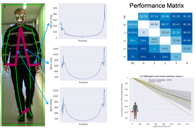
Movement Analysis
We are using sensor-free motion registrations to analyze movement patterns particularly using explainable artificial intelligence (AI) to identify the most characterizing disease and stage specific features. The application of advanced computer vision techniques allows us to discern subtle changes in motion patterns not detectable by human examiners, offering valuable diagnostic and prognostic insights. This pioneering methodology has the potential to improve the accuracy of assessments within a clinical and research setting as well as for severity monitoring at home.
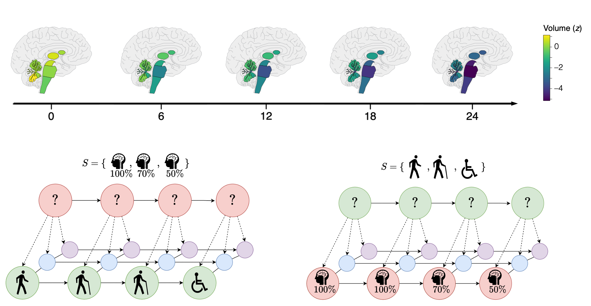
Data-Driven Disease Modeling
Data-driven disease modeling, utilizing extensive datasets allows to develop refined models that deepen our insights into ataxias and their progression. By assimilating diverse data sources such as clinical records, genetic information, and imaging data, our focus is on constructing comprehensive models that capture the complex dynamics of ataxic disorders. Our approach involves advanced statistical and machine learning techniques to identify patterns, correlations, and potential markers within the data.
Imaging Biomarkers in Ataxias
One of our main research aims is the identification of robust and reliable MR imaging biomarkers, particularly in sporadic and hereditary ataxias. Imaging biomarkers may serve as diagnostic markers, supporting early and accurate diagnosis e.g. in sporadic diseases, or as marker for progression as well as stratification, e.g. in mutation carriers close to their imminent ataxia onset. For this purpose, we combine quantitative as well as qualitative information derived from advanced MR imaging. By gaining knowledge in this field, we aim to contribute to the development of targeted interventions and personalized treatment strategies.